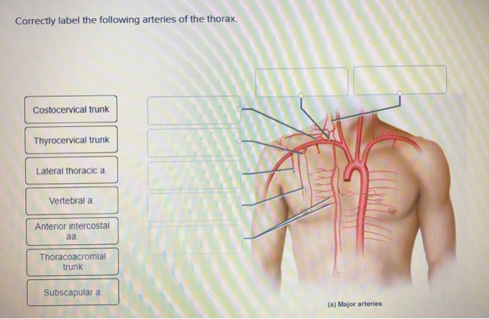
Correctly Label the Following Arteries of the Thorax
Anatomy and Physiology questions and answers. Left Superior intercostal v.

Ant Anatomy Diagram Google Search Ants Thorax Anatomy
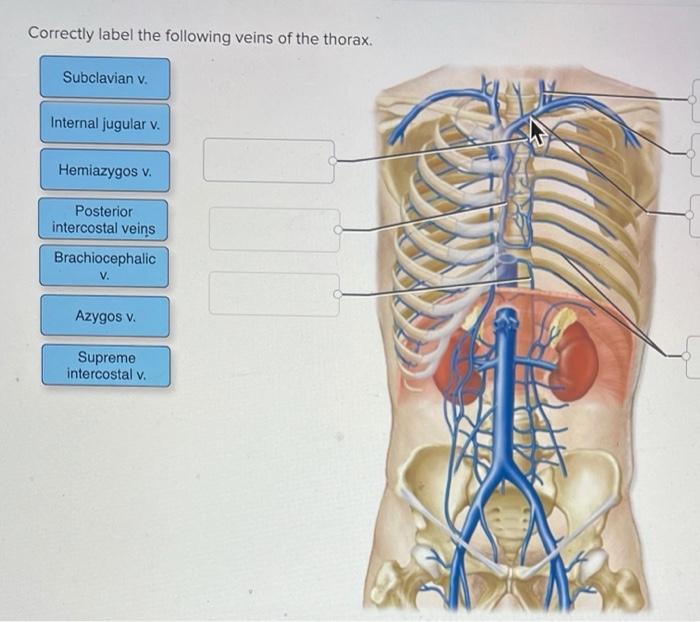
Science Anatomy and Physiology QA Library Correctly label the following veins of the thorax.

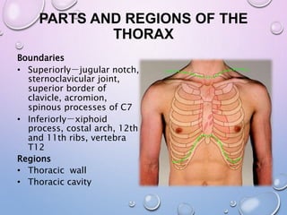
. Correctly label the following anatomical features of the thoracic cavity. Correctly label the following features of the anterior view of the thorax. Most of the arteries of the thoracic cavity arise directly from the thoracic aorta.
Correctly label the following coronary blood vessels of the heart. Correctly label the following arteries of the upper limbs. While others arise from its branchesOn the other hand the veins of the thoracic wall eventually coalesce to drain into the vena caval system.
Correctly label the following pulse points. View Screenshot 20png from BIO 1400 at CUNY Kingsborough Community College. The tunica __________ consists of smooth muscle cells arranged circularly around the blood vessel.
Correctly label the following features of the aorta and its major branches. This figure shows variations in circulatory pathways. Correctly label the following arteries of the thorax.
Theyre present only in the upper 9 spaces and every space includes 2 veins and accompanies the anterior intercostal arteries. Anatomy Tables - Arteries of the Thorax. Contributions received from several arteries vertebral posterior intercostal subcostal lumbar.
Right Superior intercostal v. The sinuatrial SA node is a patch of modified cardiomyocytes in the right atrium just under the epicardium near the superior vena cava. Blood Vessel Circulation 2020-02-18.
Categorize the following changes with regard to. 14 Aortic valve 037 Openings to coronary arteries points Skipped Left AV valve References Pulmonary valve Fibrous skeleton Right AV. Correctly label the following veins of the thorax.
Correctly label the following veins of the thorax. Arteries and veins of the thoracic wall. Correctly label the following arteries of the thorax.
Brachiocephalic trunk Radial collateral a Deep brachial a Brachial a Axillary a NA Prachiocephalic trunk Subclaviana Superior ulnar colaterala. Correctly label the pathway for the cardiac conduction system. Study quiz for Anatomy and Physiology This quiz has tags.
Correctly label the following anatomical features of the heart and thoracic cage correctly label the following vessels leading from and toward the anterior heart correctly label the following external anatomy of the posterior heart. Correctly label the following arteries of the abdomen and pelvic region. The pathway labeled _____ shows alternative routes of blood supply found in the heart.
Your Skills Rank. Anterior Intercostal Veins. They arise as multiple branches of several vessels vertebral posterior intercostal lumbar and lateral sacral aa anterior radicular aa.
Anastomose with the anterior spinal a. Anatomy and Physiology questions and answers. Correctly label the following parts of the pericardium and the heart walls.
Page 3 of 20 StudyBlue printing of Chapter 21. This is the pacemaker that initiates each. Page 12 of 20 StudyBlue printing of Chapter 21.
Correctly label the following features of the anterior view of the thorax. Coronary circulation intrinsic to the heart takes blood directly from the main artery aorta coming from the heart. This is an online quiz called Arteries of the Thorax.
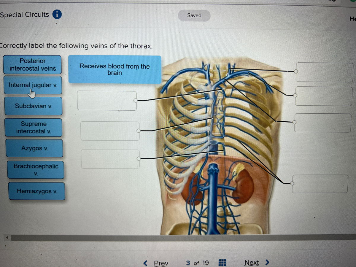
Posterior intercostal veins Receives blood from the brain Internal jugular v. Media Classify the following images into the types of vessels they represent. Posterior intercostal veins SubclaviarM Brachiocephalic V.
The thoracic wall or chest wall is a musculoskeletal structure that has a vast vascular supply. Blood Vessel Circulation 2020-02-18. Costocervical trunk Thyrocervical trunk Lateral thoracic a.
Drag each label into the appropriate position to identify the waves of a normal ECG. Correctly label the following structures related to the position of the heart in the thorax. From the quiz author.
Show transcribed image text Correctly label the following features of the anterior view of the thorax. Correctly label the great vessels that enter and exit the heart. Click on the tags below to find other quizzes on the same subject.
Special Circuits Saved Не Correctly label the following veins of the thorax. WYLLY 14 Vertebral a Anterior intercostal aa Thoracoacromial trunk Subscapular a a Major arteries. Correctly label the following veins of the thorax Subcostal v Postenor intercostal veins R ascending lumbar Lascending lumbar Lumbar veins Hemiazygos Reset Zoom.
They conclude in the upper 6 spaces they stop in the internal thoracic vein and in seventh eighth and ninth spaces they stop in the musculophrenic vein. Correctly label the following arteries of the thorax. Correctly label the following parts of the heart valves.
Correctly label the following veins of the abdomen and pelvic region. Correctly label the following major systemic arteries. Correctly label the following coronary blood vessels of the heart.

19 5 Heart Position Within The Thoracic Cavity A The Heart Is Located Within The Mediastinum Of The Thoracic Cavity Betwee Thoracic Cavity Cavities Thoracic
Solved Label The Figure With The Items Provided Obturator Chegg Com
Label The Branches Of The Aorta In This Anterior View Of The Thorax And Abdomen Thoracic Homeworklib

Chest Anatomy Human Body Sticker By Hoorahville Human Anatomy And Physiology Human Anatomy Anatomy And Physiology
![]()
Thorax Anatomy Wall Cavity Organs Neurovasculature Kenhub

Thoracic Cavity Description Anatomy Physiology Britannica

Human Ribs Google Search Human Ribs Human Body Anatomy Rib Cage Anatomy

Arteries Of The Thorax Quiz
/GettyImages-970770740-d32eb4bb2b404c95bf7243d3ce6cf51f.jpg)
Sternum Anatomy Function And Treatment
![]()
Thorax Anatomy Wall Cavity Organs Neurovasculature Kenhub

Circulatory Pathways Anatomy And Physiology Ii
![]()
Thorax Anatomy Wall Cavity Organs Neurovasculature Kenhub
:max_bytes(150000):strip_icc()/heart-and-circulatory-system-with-blood-vessels--97537745-a3bc2b2a6ca94390bfdf2696ad9bbddd.jpg)
Pulmonary Vein Anatomy Function And Significance
Solved Correctly Label The Following Arteries Of The Thorax Chegg Com

Cone Shaped Lungs Located Inside The Thorax Also Known As The Chest And Also Inside The Ribcage Are Covered In A Thin Membrane Call Lunges Rib Cage Thorax
Thorax

Heart Anatomy Anatomy And Physiology Ii
Answered Ctly Label The Following Veins Of The Bartleby
![]()
Thorax Anatomy Wall Cavity Organs Neurovasculature Kenhub


Comments
Post a Comment